

The field of restorative dentistry has shifted dramatically away from temporary, surface-level prosthetics. For individuals addressing tooth loss, basic options like removable dentures or enamel-destructive bridges have been widely replaced by a more advanced clinical standard: the dental implant. When patients research this procedure at a modern dental clinic near me, they often focus on the immediate structural components—the titanium post, the connecting abutment, and the final custom crown that restores the visual symmetry of their smile.
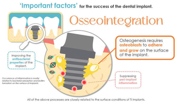
However, the true clinical success of a dental implant is not determined during the surgical appointment. The long-term stability, biting power, and permanence of the replacement tooth depend entirely on a complex biological phenomenon known as osseointegration.
Derived from the Greek osteon (bone) and the Latin integrare (to make whole), this natural cellular process represents a seamless fusion between living bone tissue and an artificial medical device. Understanding the cellular science behind this healing window helps patients understand exactly what to expect as their body works to secure their new smile.
The Discovery of Biocompatible Fusion
The scientific foundation of modern implantology was discovered by accident in the mid-twentieth century by a Swedish orthopedic surgeon named Per-Ingvar Brånemark. While conducting research on blood circulation using microscopic optical chambers enclosed in titanium sleeves anchored to animal bone tissue, he discovered something unexpected: when the study concluded, the titanium chambers could not be removed. The surrounding living bone had grown into complete contact with the metal surface, anchoring it completely.
This breakthrough proved that pure titanium is highly biocompatible. Because the human body does not recognize it as a foreign threat, it avoids triggering an adverse immune response or forming protective scar tissue. Instead, the skeletal system treats the titanium post exactly like natural bone, initiating a structural healing process that turns the medical device into a permanent part of the jaw.
The Four Phases of Osseointegration: A Biological Timeline
Once an experienced clinician at a premier dental clinic near me secures the dental implant into a precisely prepared pathway within the jaw, a multi-phase biological clock begins. This cellular journey unfolds over several months:
Phase 1: Hemostasis and Clot Formation (Minutes to Hours Post-Surgery)
The moment the implant is placed, structural microvessels within the bone tissue open, causing localized bleeding. Within minutes, the body triggers a protective clotting response (hemostasis). A dense network of fibrin proteins forms a stable mesh around the microscopic threads of the implant post. This blood clot serves as a vital biological bridge, filled with signaling proteins and growth factors that summon healing cells to the surgical site.
Phase 2: Immune Response and Cellular Infiltration (Days 1 to 3)
Specialized white blood cells (macrophages and neutrophils) migrate into the fibrin matrix. Their primary role is to sterilize the environment, clearing away microscopic tissue debris or bacteria from the surgical site. Simultaneously, these cells release chemical signals that recruit primitive mesenchymal stem cells, guiding them toward the specialized micro-texture of the titanium surface.
Phase 3: Osteogenesis and Woven Bone Formation (Weeks 1 to 4)
The recruited stem cells specialize into active bone-building cells known as osteoblasts. These cells begin laying down a framework of collagen fibers, which quickly mineralize to form early “woven bone.” This initial bone structure grows directly into the microscopic pores and acid-etched grooves of the implant post. By the end of the first month, this intricate bone network provides initial stabilization, anchoring the post within the jaw.
Phase 4: Lamellar Bone Remodeling (Months 3 to 6)
Woven bone is formed rapidly, but it lacks the organized, dense architecture required to withstand the high mechanical forces of daily chewing. Over the next several months, a continuous structural update occurs. Specialized bone-resorbing cells (osteoclasts) break down the temporary woven bone, while osteoblasts replace it with highly organized, load-bearing lamellar bone. This mature bone layer aligns precisely along the stress lines of the implant, providing the deep structural strength needed to support the final crown.
Structural Progression: Primary vs. Secondary Stability
During this multi-month biological timeline, the implant experiences a complex transition in how it is stabilized within the jawbone. This path is tracked carefully by clinicians using specialized electronic resonance metrics.
| Evaluation Phase | Primary Mechanical Stability | Secondary Biological Stability |
| Source of Support | Physical friction between the implant threads and the bone wall | New living bone matrices grown directly into the surface pores |
| Peak Timeline | Immediately post-op (Day 0) | Progressively builds from Week 3 to Month 6 |
| Tissue Type | Pre-existing cortical and trabecular jawbone | Mature, load-bearing lamellar bone |
| Clinical Vulnerability | Declines over the first 2 weeks as old bone relaxes | Highly resilient; grows stronger under controlled biting pressure |
The transition period between Week 2 and Week 4 is often referred to by implantologists as the stability dip. This occurs because the initial mechanical friction decreases slightly before the new biological bone has fully mineralized. This crucial window explains why providers place patients on a soft-food diet during early recovery, shielding the implant from heavy forces while the body works to build its biological anchor.
Protecting the Integration: The Value of Local Follow-Up Care
Because the cellular steps of osseointegration are deeply impacted by a patient’s systemic health and daily habits, staying aligned with an accessible dental clinic near me is essential for long-term success.
- Managing Early Inflammation: If oral bacteria slip past the gum line during the early healing phases, they can trigger an inflammatory condition known as peri-implant mucositis. Left unmanaged, this can progress to bone loss around the post, disrupting the integration process before it finishes. Regular, local follow-ups allow the clinical team to screen the gums and keep the site sterile.
- Bite Forces and Alignment: Once the bone has fully integrated, the custom crown is attached. Having your care handled by a nearby clinic ensures that any tiny bite imbalances can be adjusted instantly. A tooth that hits too hard can overload the new bone matrix, causing micro-fractures in the surrounding tissue that could lead to implant failure.
- Lifestyle and Wellness Tracking: Medical history, direct micro-circulation health, and habits like tobacco use profoundly alter the body’s healing speed. Local specialists work closely with patients to manage these health variables, ensuring the jawbone receives the steady blood supply and nutrients it needs to complete the integration process safely.
Conclusion: The Triumph of Biological Engineering
The remarkable success rate of modern dental implant dentistry is a direct testament to the body’s natural ability to adapt and heal. A dental implant does not simply sit inside the mouth like a traditional bridge or removable denture; it systematically integrates with the human skeletal system at a cellular level.
By understanding the intricate timeline of osseointegration—from the initial blood clot to the creation of mature, load-bearing bone—patients can appreciate the scientific care that guides each step of their recovery. By pairing this advanced biological process with the diagnostic tools and attentive follow-up care of a premier dental clinic near me, individuals can step forward with absolute confidence, knowing their new smile is anchored by a permanent, scientifically proven foundation built to last a lifetime.